
Ensembles de lames de microscope pour la salle de classe

Invitrogen™ Kit de coloration pour la prolifération des neurites

Le Molecular Probes™ kit de coloration de la prolifération des neurites permet une mesure rapide et simple de la prolifération des neurites et de la viabilité cellulaire dans le même échantillon.


Eisco™ Racine de coctotyledone, coupe transversale
Lame de microscope préparée d’une coupe transversale de la racine monocotylédonée et dicotylédone. Cette lame combinée offre une comparaison facile des stuctures racinaires caractéristiques d’un monocotyledon et d’un dicotyledône typiques.
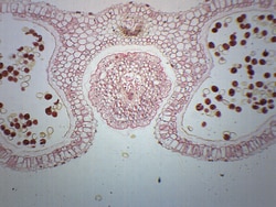
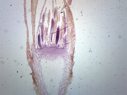
Eisco™ Antère de lilium, section transversale
Lame de microscope préparée d’une coupe transversale d’une anthère de lilium mature contenant du pollen. Anther est une partie de la fleur où le pollen est produit. Montrant des structures caractéristiques de l’anthère de lilium et du pollen. Excellent choix pour les salles de biologie.

Eisco™ Tilia 1 an de tige, section transversale
Lame de microscope préparée d’une section transversale de tilia vieille d’un an. Le tilia, communément appelé tilleul, est un dicothés. Montrant les structures caractéristiques d’une tige dicothéstylé. C’est un excellent choix pour les classes de biologie afin d’explorer les relations structure-fonction selon les normes NGSS.

Eisco™ Section de jéjunum (mammifère)
Lame de microscope préparée de jéjunum de mammifères. Le jéjunum fait partie de l’intestin grêle. Coloré pour une meilleure visualisation des couches caractéristiques de l’intestin grêle. Excellent choix pour les cours de biologie qui étudient le système digestif.

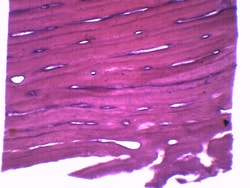
Eisco™ Lame de microscope préparée - Muscle squelettique humain, section logitudinale
Lame de microscope préparée du muscle squelettique humain

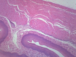
Eisco™ Lame de microscope préparée - Artère et veine moyennes, section transversale
Lame de microscope préparée d’une coupe transversale de l’artère et de la veine moyennes

Eisco™ Labame de microscope préparée - Frottis sanguin humain, tache d’H &E
Lame de microscope préparée d’un frottis sanguin humain
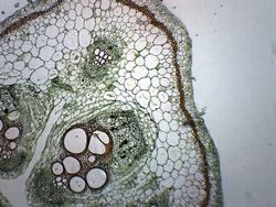
Eisco™ Tige de citrouille, section transversale combinée et section longitudinale
Lame de microscope préparée d’une tige de citrouille. Cette diapositive montre les sections longitudinales et transversales combinées, permettant une meilleure visualisation et compréhension des structures de tiges.

Eisco™ Muscle cardiaque, mammifères, section transversale et section longitudinale
Lames de microscope préparées du muscle cardiaque des mammifères. La combinaison de la section longitudinale et de la section transversale permet une meilleure visualisation et compréhension de la morphologie et de l’anatomie du muscle cardiaque.
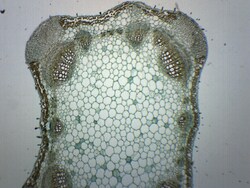
Eisco™ Tige lavande, coupe transversale
Lame de microscope préparée de tige de lavande. Section transversale. La lavande est un dicotyglottes. Présente les structures générales d’une tige dicothéstylétine typique. Idéal pour les classes de biologie afin d’explorer les relations structure-fonction selon les normes NGSS.
Eisco™ Épithélium de l’œsophage, section transversale
Lame de microscope préparée d’une coupe transversale de l’œsophage, montre des cellules épithéliales. L’œsophage est tapissé de cellules épithéliales squameuses. Excellent outil pour les cours de biologie.
Eisco™ Moss Archegonia, section longitudinale
Lame de microscope préparée d’une section lontitudinale d’archegonies de mousse. Les archégonies sont des organes qui produisent et contiennent des ovules ou gamètes femelles. Excellent choix pour les cours de biologie étudiant la reproduction sexuelle des plantes et des champignons.
Eisco™ Os, section transversale et section longitudinale
Une lame composite préparée de la section transversale et longitudinale d’un os. Coloré pour une meilleure visualisation des structures caractéristiques. Idéal pour les classes de biologie afin d’explorer les relations structure-fonction selon les normes NGSS.