
Classroom Microscope Slide Sets

Invitrogen™ Neurite Outgrowth Staining Kit

The Molecular Probes™ Neurite Outgrowth Staining Kit allows for quick and simple measurement of neurite outgrowth and cell viability in the same sample.

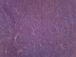
Eisco™ Spleen Section - Mammalian, H&E Stain
Prepared microscope slide of spleen section stained with Hematoxylin and Eosin. Spleen is a lymphatic tissue. Shows general structures.

Eisco™ Tilia 1 Yr Stem, Cross Section
Prepared microscope slide of one year old tilia stem cross section. Tilia, commonly known as basswood is a dicot. Showing characteristic structures of a dicot stem. Great choice for biology classrooms to explore structure function relationships as per NGSS standards.
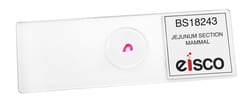
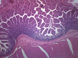
Eisco™ Jejunum Section (Mammal)
Prepared microscope slide of mammalian jejunum. Jejunum is a part of small intestine. Stained for better visualization of characteristic layers of small intestines. Great choice for biology classrooms studying digestive sysem.

Eisco™ Blood Smear (Bird), GS Stain
Prepared microscope slide of bird blood smear stained with Giemsa stain. Showing characteristic bird blood cells and their relative ratios. Great choice for biology classrooms to explore structure function relationships as per NGSS standards.
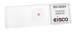
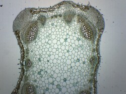
Eisco™ Tilia Old Stem (Basswood), Cross Section
Prepared microscope slide of Tilia old stem. Tilia is commonly known as basswood. Stained for better visualization of characteristic structures, including secondary xylem annual rings. Great choice for biology classrooms to explore structure function relationships as per NGSS standards.
Eisco™ Sperm Smear, Mammalian
Prepared microscope slide of mammalian sperm smear. Shows characteristic features of sperm cells. Ideal choice for biology classrooms or curious scientists.
Eisco™ Monocot Leaf Cross Section, General Structures
Prepared microscope slide of a cross section or a monocot leaf. Leaves are important plant organs where photosynthesis and water exchange takes place. This slide shows general structures of a typical monocot leaf.
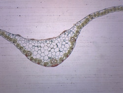
Eisco™ Lavender Stem, Cross Section
Prepared microscope slide of lavender stem. Cross section. Lavender is a dicot. Shows general structures of a typical dicot stem. Ideal for biology classrooms to explore structure function relationships as per NGSS standards.
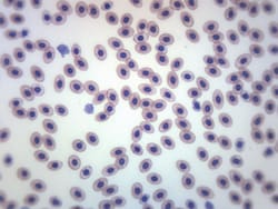
Eisco™ Frog Blood Smear, GS Stain
Prepared microscope slide of frog blood smear. An example of blood cells from a cold-blooded animal. Stained with GS stain for better visualisation. Whole Mount.
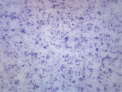
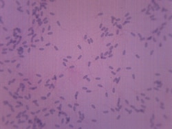
Eisco™ Bacillus - Mixed, Gram Positive and Negative
Prepared microscope slide of mixed Bacillus culture, gram positive and gram negative. Ideal for studying rod-shaped morphology. Gram positive bacteria appear purple, as they are stained with crystal violet stain. Gram-negative bacillus is stained with carbol fuchsin and appear the color of fuchsia.
Eisco™ Colon (Mammal), Cross Section
Prepared microscope slide of a cross section of a mammalian colon. Stained for better visualization of characteristic structures and tissues. Great choice for biology classrooms to explore structure function relationships as per NGSS standards.
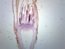
Eisco™ Moss Archegonia, Longitudinal Section
Prepared microscope slide of a lontitudinal section of moss archegonia. Archegonia are organs which produce and contain ova or female gametes. Great choice for biology classrooms studying sexual reproduction of plants and fungi.
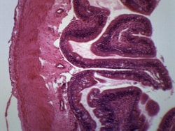
Eisco™ Frog Intestine, Cross Sectin and H&E Stain
Prepared microscope slide of a cross section of frog intestine. Stained with hematoxylin and eosin for better visualization of characteristic structures and tissue layers. Great choice for biology classrooms to explore structure function relationships as per NGSS standards.