
Classroom Microscope Slide Sets

Invitrogen™ Neurite Outgrowth Staining Kit

The Molecular Probes™ Neurite Outgrowth Staining Kit allows for quick and simple measurement of neurite outgrowth and cell viability in the same sample.


Rocks and Minerals Slide Sets
Identify forms, colors, refraction and fossil inclusions.
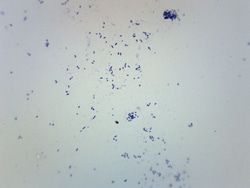
Eisco™ Streptococcus Pneumoniae, Gram Positive
Prepared microscope slide of Streptococcus pneumoniae, stained with Gram stain. Streptococcus pneumoniae walls retain the Crystal Violet in the Gram stain,thus Gram-positive and violet in appearance. Shows characteristic size and shape of Streptococcus pneumoniae.

Representative Protist Set
All four classes of protist are represented.

United Scientific™ Prepared Slide Set, 4 Slides
Set offers a variety of different prepared slides for classroom use.
.JPG-250.jpg)
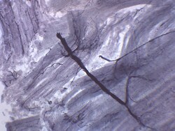
Eisco™ Bacillus Subtilis, Gram Positive
Prepared microscope slide of Bacillus Subtillis. Wholemount. Bacillus Subtillis is a gram-positive bacteria found in soil and gastro-intestinal tract of humans. Ideal addition to a microbiology slide collection.

Plant Cell Slide Set
Plant tissues from root tip to flower parts.
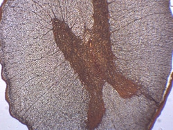
Eisco™ Fern Life Cycle, Composite
Prepared composite microscope slide of fern life cycle. The slide shows 3 stages of fern life cycle: Gemetophyte, Young Sporophyte, and Sporangia.
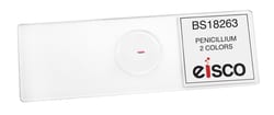
Eisco™ Penicillium
Prepared microscope slide of Penicillium. Penicillium is a genus of fungi, some members of which produce penicillin. Showing characteristic morphology. Great choice for biology classrooms.
Eisco™ Prepared Microscope Slide - Motor Nerve Ending Plates, Whole Mount
Prepared microscope slide of motor nerve ending plates
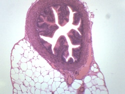
Eisco™ Prepared Microscope Slide - Spinal Cord Cross Section
Prepared microscope slide of spinal cord

Eisco™ Prepared Microscope Slide - Skin Hairy Section, H&E Stain
Prepared microscope slide of hairy section of skin
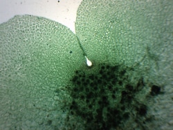
Eisco™ Nostoc Cross Section, FS & FG Stain
Prepared microscope slide of Nostoc cross section, stained with FS and FG stain. Nostoc is a genus of cyanobacteria that forms colonies in a gelatinous sheath. Ideal addition to a microbiology collection.
Eisco™ Fallopian Tube - Mammalian, Cross Section
Prepared microscope slide of a cross section of a Fallopian Tube. The Fallopian tube is a part of the female reproductive system. Shows general structures.