
Classroom Microscope Slide Sets

Invitrogen™ Neurite Outgrowth Staining Kit

The Molecular Probes™ Neurite Outgrowth Staining Kit allows for quick and simple measurement of neurite outgrowth and cell viability in the same sample.


Eisco™ Excretory System Ureter, Cross Section
Prepared microscope of a cross section of mammalian ureter, a part of excretory system. Stained to show characteristic tissues, including transitional epithelium. Transitional epithelium is comprised of two or more layers of cuboidal or columnar cells. Great for biology classrooms.

Eisco™ Prepared Microscope Slide - Typical Bacteria
Prepared microscope slide of typical bacteria

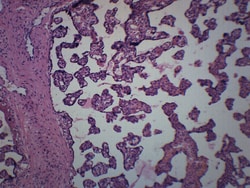
Eisco™ Endocrine System, Thyroid Gland
Prepared microscope slide of mammalian thyroid gland. Thyroid gland is a part of endocrine system. Shows characteristic structures and components of the thyroid.
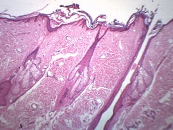
Eisco™ Hair Follicle
Prepared microscope slide of hair follicle. Showing hair follicles, epidermis, and other skin structures. Great choice for biology classrooms to explore structure function relationships as per NGSS standards.
Eisco™ Anabaena, Wholemount
Prepared microscope slide of Anabaena, wholemount. Anabaena is a flilamentous cyanobacteria commonly found as plankton. It is best known for its nitrogen-fixing abilities and forms symbiotic relationships with certain plants. Ideal for studying ecology.
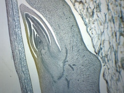
Eisco™ Zea Mays Corn Grain, Longitudinal Section
Prepared microscope slide of Zea Mays Corn grain. Longitudinal section. Stained to show different cell types present in corn seed. Excellent choice for biology classrooms to explore structure-function relationships as per NGSS standards.
Eisco™ Placenta - Human, Section
Prepared microscope slide of a section of human placenta. Stained for better visualization of characteristic structures. Ideal for biology classrooms to explore structure function relationships as per NGSS standards.
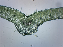
Eisco™ Ligustrum Leaf, Cross Section
Prepared microscope slide of a cross section of a Ligustrum leaf. Leaves are important organs of the plant, where photosynthesis and gas and water exchange takes place. Excellent tool for biology classrooms to explore structure-function relationships as per NGSS standards.
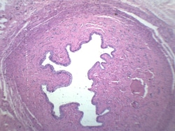
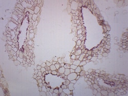
Eisco™ Urethra - Mammalian, Cross Section
Prepared microscope slide of a cross section of mammalian female Urethra. The Urethra is a part of the urinary/renal system. Stained to show general structures.
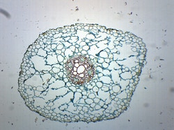
Eisco™ Ranunculus Root, Cross Section
Prepared microscope slide of a cross section of a ranunculus root. Ranunculus, commonly known as buttercup, is a dicot. Showing typical structures of a dicot root. Great choice for biology classrooms to explore structure function relationships as per NGSS standards.

General Biology Slide Set A
Prepared slides of animal and plant structures in whole mount (w.m.), longitudinal section (l.s.) and cross-section (c.s.) preparations.

Eisco™ Prepared Microscope Slide - Pseudostratified Columnar Ciliated Epithelium, H&E Stain
Prepared microscope slide of Pseudostratified Columnar Ciliated Epithelium

Eisco™ Tilia 1 Yr Stem, Cross Section
Prepared microscope slide of one year old tilia stem cross section. Tilia, commonly known as basswood is a dicot. Showing characteristic structures of a dicot stem. Great choice for biology classrooms to explore structure function relationships as per NGSS standards.