
Immunohistochemistry Kits and Reagents


Invitrogen™ Alexa Fluor™ 488 Tyramide SuperBoost™ Kit, streptavidin

SuperBoost™ tyramide signal amplification is the most sensitive method for detection of low abundant targets in multiplexable fluorescent immunocytochemistry (ICC), immunohistochemistry (IHC ), and in situ hybridization (ISH).

Invitrogen™ Predictor™ hERG Fluorescence Polarization Assay Kit
Predictor™ hERG Fluorescence Polarization Assay Kit

Thermo Fisher Scientific™ ReadyProbes™ Hydrophobic Barrier Pap Pen
The ReadyProbes Hydrophobic Barrier Pap Pen creates a water-repellent barrier when used on a microscope slide or coverslip.

Invitrogen™ Alexa Fluor™ 647 Tyramide SuperBoost™ Kit, goat anti-rabbit IgG
Features poly-HRP-conjugated goat anti-rabbit IgG secondary antibody. Alexa Fluor™ 647 Tyramide SuperBoost™ Kit combine brightness of AlexaFluor dyes with high signal amplification of a poly-HRP-mediated tyramide labeling reaction to produce s
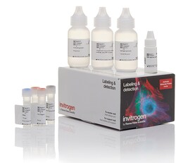
Invitrogen™ Biotin XX Tyramide SuperBoost™ Kit, Goat anti-Mouse IgG
SuperBoost™ tyramide signal amplification is the most sensitive method for detection of low abundant targets in multiplexable fluorescent immunocytochemistry (ICC), immunohistochemistry (IHC ), and in situ hybridization (ISH).

MilliporeSigma™ Calbiochem™ IgG from Rabbit Plasma
Used as negative control with specific rabbit IgG-type primary antibodies.

Invitrogen™ eBioscience™ IHC Antigen Retrieval Solution - Low pH (10X)
Designed for use during the heat induced epitope retrieval (HIER) step prior to immunohistochemistry on formalin-fixed paraffin-embedded tissue sections. Used in combination with heat has been shown to restore the antigenicity of proteins modified during the formalin fixation of tissue.

Thermo Scientific™ Pierce™ Peroxidase IHC Detection Kit
Mouse Monoclonal Antibody
Invitrogen™ eBioscience™ DAB Advanced Chromogenic Kit
Contains the necessary reagents to prepare a DAB substrate working solution.
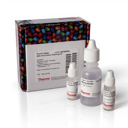
Thermo Scientific™ ABC Peroxidase Standard Staining Kit
Detect biotinylated secondary antibodies by IHC using the highly sensitive avidin-biotin complex (ABC) technique.

Thermo Scientific™ Metal Enhanced DAB Substrate Kit
Special formulation of diaminobenzidine peroxidase substrate that uses cobalt and nickel chloride to yield intense color for IHC staining.

Invitrogen™ eBioscience™ IHC Antigen Retrieval Solution - High pH (10X)
Designed for use during the heat induced epitope retrieval (HIER) step prior to immunohistochemistry on formalin-fixed paraffin embedded tissue sections. Use in combination with heat has been shown to restore the antigenicity of proteins modified during the formalin fixation of tissue.

Invitrogen™ ReadyProbes™ Tissue Autofluorescence Quenching Kit
Minimizes unwanted autofluorescence in FFPE and frozen tissues due to aldehyde fixation, red blood cells, and structural elements such as collagen and elastin

Invitrogen™ Alexa Fluor™ 647 Tyramide SuperBoost™ Kit, goat anti-mouse IgG
Features poly-HRP-conjugated goat anti-mouse IgG secondary antibody. Alexa Fluor™ 647 Tyramide SuperBoost™ Kit combine brightness of AlexaFluor dyes with high signal amplification of a poly-HRP-mediated tyramide labeling reaction to produce se
Invitrogen™ eBioscience™ BrdU Kit for IHC/ICC Colorimetric
Contains the necessary reagents and buffers for identifying and examining proliferating cells by immunohistochemical or immunocytochemical analysis. Optimized for IHC with both frozen and paraffin-embedded BrdU-labeled mouse intestine and ICC of BrdU-pulsed HeLa cells grown on culture slides.