
Ensembles de lames de microscope pour la salle de classe

Invitrogen™ Kit de coloration pour la prolifération des neurites

Le Molecular Probes™ kit de coloration de la prolifération des neurites permet une mesure rapide et simple de la prolifération des neurites et de la viabilité cellulaire dans le même échantillon.

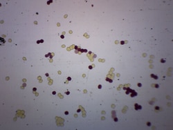
Lames d’histologie du sang animal
Étudiez les groupes sanguins des animaux.

Lames préparées pour les racines et les tiges
Les diapositives offrent une vue agrandie des gymnospermes et angiospermes pour une étude détaillée.

United Scientific™ Jeu de diapositives préparé par la biologie, 13 diapositives
Set offre une variété de diapositives préparées différentes pour une utilisation en classe.

Eisco™ Labame de microscope préparée - Section transversale de tige monocotiledonée
Lame de microscope préparée de la section transversale de la tige monocotiledonée

Eisco™ Lame de microscope préparée - Section de tissu aréolaire
Lame de microscope préparée de la section de tissu aréolaire

Eisco™ Lame de microscope préparée - Sarcodina amieba Proteus, montage complet
Lame de microscope préparée d’amibe

Eisco™ Lame de microscope préparée - Système endocrinien - Glande surrénale
Lame microscopique préparée de la glande surrénale
Eisco™ Muscle lisse, section longtitudinale, mammifère
Lame de microscope préparée du muscle lisse des mammifères. Section longtitudinale. Idéal pour les classes de biologie souhaitant explorer la connexion structure-fonction selon les normes NGSS.
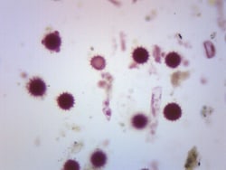
Eisco™ Comparaison des Dicot Pollens, Wholemount
Lame de microscope préparée d’un mélange de pollens de dicothéthéres. Wholemount. Idéal pour les classes de biologie souhaitant explorer la connexion structure-fonction selon les normes NGSS.
.JPG-250.jpg)
Eisco™ Conjugaison Protista Paramecium, Wholemount
Lame de microscope préparée de la conjugaison de Paramecium. Wholemount. Paramecium est un organisme unicellulaire que l’on trouve principalement en milieu d’eau douce. Cet eucaryote appartient au genre des protozoaires cilliés. Cette diapositive montre la reproduction du paramécium - conjugaison.

Eisco™ Clostridium botulinum, Gram positif
Lame de microscope préparée de Clostridium botulinum à gram positif. Wholemount. Clostridium Botulinum est une bactérie en forme de bâtonnet, communément connue pour produire de la toxine botulinique. Idéal pour une collecte de lames en microbiologie.
Eisco™ Œuf d’Ascaris Lumbricoides, Wholemount
Lame de microscope préparée de l’œuf d’Ascaris Lumbircoides. Ascaris Lumbricoides est un gros ver rond parasite que l’on trouve dans les intestins humains. Wholemount
.JPG-250.jpg)
Eisco™ Extrémité de la racine de lilium, section transversale
Lame de microscope préparée de la section transversale de la pointe de la racine de Lilium. La pointe de la racine est le site de croissance d’une plante, et donc un lieu où se produit la mitose. Outil idéal pour étudier les structures des extrémités racinaires ainsi que la mitose.