
Classroom Microscope Slide Sets


United Scientific Supplies Prepared General Biology Slides, Set of 25
Set of 25 general biology slides comes with a case for easy storage and retrieval.
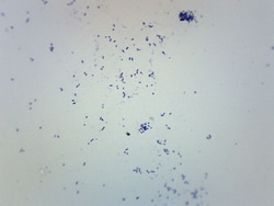
Eisco™ Streptococcus Pneumoniae, Gram Positive
Prepared microscope slide of Streptococcus pneumoniae, stained with Gram stain. Streptococcus pneumoniae walls retain the Crystal Violet in the Gram stain,thus Gram-positive and violet in appearance. Shows characteristic size and shape of Streptococcus pneumoniae.

Histology of Mammalia, Basic Set
Precision slides are mounted by expert microtechnicians.
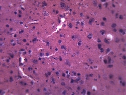
Eisco™ Brain - Mammalian, Composite Section
Prepared composite microscope slide of mammalian brain tissue. Characteristic structures are clearly identifiable. Ideal for biology classrooms studying nervous system.
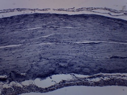
Eisco™ Striated Skeletal Muscle (Frog)
Prepared microscope slide of frog striated skeletal muscle. Stained for better visualization of characteristic morphology and structures. Great choice for biology classrooms to explore structure function relationships as per NGSS standards.
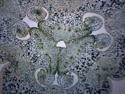
Eisco™ Lilium Ovary
Prepared microscope slide of lilium ovary. Stained for better visualization of characteristic structures including ovules and embryo sacs. Great choice for biology classrooms to explore structure function relationships as per NGSS standards.
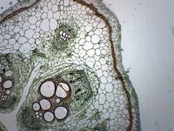
Eisco™ Pumpkin Stem, Combined Cross Section and Longitudinal Section
Prepared microscope slide of a Pumpkin stem. This slide shows combined longitudinal and cross sections, allowing better viewing and understanding of stem structures.

Histology of Mammalia, Advanced Set
High-quality set with 50 more slides for broader look.

United Scientific™ Biology Prepared Slide Set, 12 Slides
Set offers a variety of different prepared slides for classroom use.

Fisher Science Education™ Animal Cells Basic Slide Set
A variety of basic animal cell slides.


Endocrine System Slide Set
A microscopic view of the endocrine system.
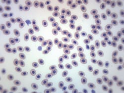
Eisco™ Frog Blood Smear, GS Stain
Prepared microscope slide of frog blood smear. An example of blood cells from a cold-blooded animal. Stained with GS stain for better visualisation. Whole Mount.

Eisco™ Frog Skin Section
Prepared microscope slide of a section of frog skin. Showing the muscle, dermis and epidermis layers as well as characteristic structures such as mucus glands, blood vessels and chormatophores. Great choice for biology classrooms.

Eisco™ Salix Branch Root Origin, Cross Section
Prepared microscope slide of a section of salix branch root origin. Stained for easy visualization of the origin of the root in a new branch cutting of a willow. Showing characteristic structures. Great choice for biology classrooms.