
Classroom Microscope Slide Sets

Invitrogen™ Neurite Outgrowth Staining Kit

The Molecular Probes™ Neurite Outgrowth Staining Kit allows for quick and simple measurement of neurite outgrowth and cell viability in the same sample.

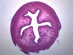
Eisco™ Uterus Section (Animal)
Prepared microscope slide of animal uterus section. Uterus is a part of reproductive system. Stained for better visualization of characteristic layers and structures. Great choice for biology classrooms to explore structure function relationships as per NGSS standards.

Eisco™ Monocot Root General Structures, Cross Section
Prepared microsocpe slide of a cross section of a monocot root. Stained for better visualization of general structures. Great choice for biology classrooms to explore structure function relationships as per NGSS standards.

Eisco™ Herbaceous Roots, Cross Section
Prepared microscope slide of a cross section of herbaceous roots. Stained for better visualization of characteristic structures. Great choice for biology classrooms to explore structure function relationships as per NGSS standards.
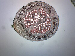
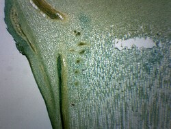
Eisco™ Pine Single Needle Leaf, Cross Section
Prepared microscope slide of a cross section of a single pine needle leaf. Stained for better visualization of characteristic structures. Great choice for biology classrooms to explore structure function relationships as per NGSS standards.
Eisco™ Old & Young Dicot Roots, Cross Section
Prepared composite slide of a cross section of old and young dicot root. Old and young dicot root cross sections are placed side by side for convenient comparison. Showing characteristic structures of dicot root. Great choice for biology classrooms.
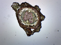
Eisco™ Young Pine Root, Cross Section
Prepared microscope slide of a cross section of young pine root. Stained for better visualization of characteristic structures. Great choice for biology classrooms to explore structure function relationships as per NGSS standards.
Eisco™ Pine Meristematic Stem, Cross Section
Prepared microscope slide of a cross section of pine meristematic stem. Stained for better visualization of characteristic structures. Great choice for biology classrooms to explore structure function relationships as per NGSS standards.
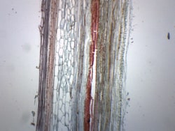
Eisco™ Pumpkin Stem (Cucurbita), Logitudinal Section
Prepared microscope slide of a longtitudinal section of pumpkin stem. Cucurbita or pumpkin is a dicot. Showing characteristic structures of dicot stems. Great choice for biology classrooms to explore structure function relationships as per NGSS standards.
Eisco™ Gymnosperm Angiosperm Stem
Prepared microscope slide of gymnosperm and angiosperm stem section. Angiosperms are plants which have flowers and fruits, while gymnosperms have no flowers and fruits and have unenclosed seeds. Great choice for biology classrooms to examine the stems of gymnosperms and angiosperms side by side.
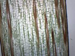
Eisco™ Zea Mays Stem, Longitudinal Section
Prepared microscope slide of a longtitudinal section of zea mays stem. Zea mays, commonly known as corn is a monocot. Stained for better visualization of characteristic monocot structures. Great choice for biology classrooms to explore structure function relationships as per NGSS standards.
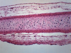
Eisco™ Frog Cartilage Section
Prepared microscope slide of a section of frog cartilage. Stained for better visualization of characteristic morphology and structures. Great choice for biology classrooms to explore structure function relationships as per NGSS standards.
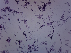
Eisco™ Azotobacter, Gram Negative
Prepared microscope slide of azotobacter. Stained with Gram stain. Azotobacter species are Gram-negative bacteria commonly found in water, soils, and plants. Showing characteristic morphology. Great choice for biology classrooms.

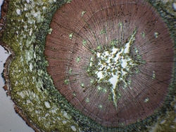
Eisco™ Tilia 2 Yr Stem (Basswood), Cross Section
Prepared microscope slide of a cross section of two year old tilia stem. Tilia, commonly known as basswood, is a dicot. Stained for better visualization of characteristic structures including secondary xylem. Great choice for biology classrooms to explore structure function relationships.