
Classroom Microscope Slide Sets

Invitrogen™ Neurite Outgrowth Staining Kit

The Molecular Probes™ Neurite Outgrowth Staining Kit allows for quick and simple measurement of neurite outgrowth and cell viability in the same sample.

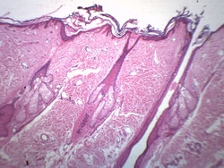
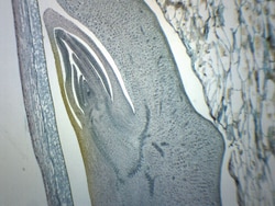
Eisco™ Hair Follicle
Prepared microscope slide of hair follicle. Showing hair follicles, epidermis, and other skin structures. Great choice for biology classrooms to explore structure function relationships as per NGSS standards.
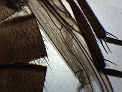
Eisco™ Musca Domestica, Whole Mount
Prepared microscope slide of musca domestica, whole mount. Shows general structures. Great choice for biology classrooms to explore structure-function relationships as per NGSS standards.
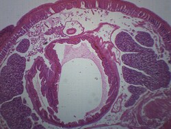
Eisco™ Annelids (Earthworm), Cross Section
Prepared microscope slide of a cross section of an earthworm. Stained to clearly show characteristic structures. Ideal for biology classrooms to explore structure function relationships as per NGSS standards.


Fisher Science Education™ Animal Cells Basic Slide Set
A variety of basic animal cell slides.
Eisco™ Anabaena, Wholemount
Prepared microscope slide of Anabaena, wholemount. Anabaena is a flilamentous cyanobacteria commonly found as plankton. It is best known for its nitrogen-fixing abilities and forms symbiotic relationships with certain plants. Ideal for studying ecology.
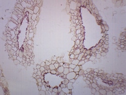
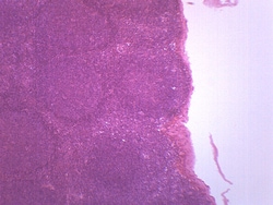
Eisco™ Zea Mays Corn Grain, Longitudinal Section
Prepared microscope slide of Zea Mays Corn grain. Longitudinal section. Stained to show different cell types present in corn seed. Excellent choice for biology classrooms to explore structure-function relationships as per NGSS standards.
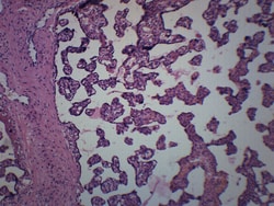
Eisco™ Placenta - Human, Section
Prepared microscope slide of a section of human placenta. Stained for better visualization of characteristic structures. Ideal for biology classrooms to explore structure function relationships as per NGSS standards.

Representative Algae Set
Observe and compare various algae.
Eisco™ Mycobacterium Tuberculosis
Prepared microscope slide of mycobacterium tuberculosis, wholemount. Mycobacterium tuberculosis is a gram positive pathogenic bacteria causing tuberculosis in humans. Ideal for microbiology studies at home or in the classroom.
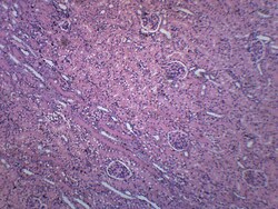
Eisco™ Kidney Section (Human), Histological Structures
Prepared microscope slide of a section of human kidney. Showing characteristic histological structures. Great choice for biology classrooms to explore structure function relationships as per NGSS standards.
Botany Survey Set
This 25-slide set ranges from bacteria through higher plants.
Eisco™ Fasciola Hepatica, Whole Mount
Prepared microscope slide of Fasciola Hepatica, commonly known as a liver fluke. This flatworm is a parasitic trematode that infects livers of mammals. Whole Mount. Shows general structures.