
Ensembles de lames de microscope pour la salle de classe


United Scientific Supplies Diapositives de biologie générale préparées, ensemble de 25
Un ensemble de 25 diapositives de biologie générale est livré avec un boîtier pour un stockage et une récupération faciles.
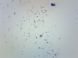
Eisco™ Streptococcus pneumoniae, Gram positif
Lame de microscope préparée de Streptococcus pneumoniae, colorée à la coloration de Gram. Les parois de Streptococcus pneumoniae conservent le violet cristallin dans la coloration de Gram, ce qui donne une apparence Gram-positive et violette. Présente la taille et la forme caractéristiques de Streptococcus pneumoniae.

Histologie des mammifères, ensemble de base
Les lames de précision sont montées par des microtechniciens experts.
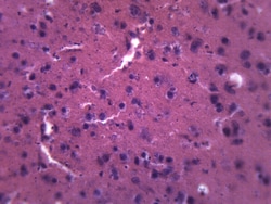
Eisco™ Cerveau - Mammifère, section composite
Lame de microscope composite préparée de tissu cérébral de mammifères. Les structures caractéristiques sont clairement identifiables. Idéal pour les classes de biologie étudiant le système nerveux.
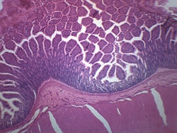
Eisco™ Côlon (mammifère), section transversale
Lame de microscope préparée d’une coupe transversale d’un côlon mammifère. Coloré pour une meilleure visualisation des structures et tissus caractéristiques. C’est un excellent choix pour les classes de biologie afin d’explorer les relations structure-fonction selon les normes NGSS.
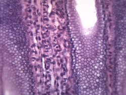
Eisco™ Cuboïdal simple, épithélium
Lame de microscope préparée d’épithélium cuboïdal simple. L’épithélium cuboïdal simple se trouve dans les organes sécréteurs et les parois des bronchioles respiratoires. Idéal pour les classes de biologie étudiant l’anatomie et examinant les liens structure-fonction selon les normes NGSS.
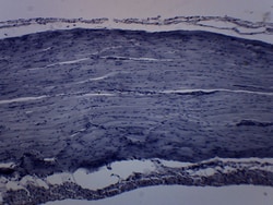
Eisco™ Muscle squelettique strié (grenouille)
Lame de microscope préparée du muscle squelettique strié de grenouille. Coloré pour une meilleure visualisation de la morphologie et des structures caractéristiques. C’est un excellent choix pour les classes de biologie afin d’explorer les relations structure-fonction selon les normes NGSS.
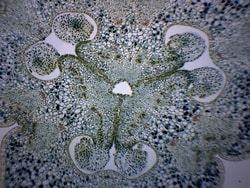
Eisco™ Ovaire de lilium
Lame de microscope préparée de l’ovaire lilium. Coloré pour une meilleure visualisation des structures caractéristiques, y compris les ovules et les sacs embryonnaires. C’est un excellent choix pour les classes de biologie afin d’explorer les relations structure-fonction selon les normes NGSS.
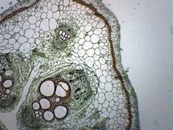
Eisco™ Tige de citrouille, section transversale combinée et section longitudinale
Lame de microscope préparée d’une tige de citrouille. Cette diapositive montre les sections longitudinales et transversales combinées, permettant une meilleure visualisation et compréhension des structures de tiges.

Histologie des mammifères, ensemble avancé
Un ensemble de haute qualité avec 50 diapositives supplémentaires pour un look plus large.

United Scientific™ Jeu de diapositives préparé par la biologie, 12 diapositives
Set offre une variété de diapositives préparées différentes pour une utilisation en classe.

Fisher Science Education™ Jeu de diapositives de base sur les cellules animales
Une variété de lames de cellules animales de base.


Jeu de diapositives du système endocrinien
Une vue microscopique du système endocrinien.
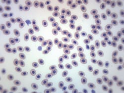
Eisco™ Tache de sang de grenouille, tache GS
Lame de microscope préparée d’un frottis de sang de grenouille. Un exemple de cellules sanguines provenant d’un animal à sang froid. Teinté avec une teinture GS pour une meilleure visualisation. Toute la monture.