Learn More
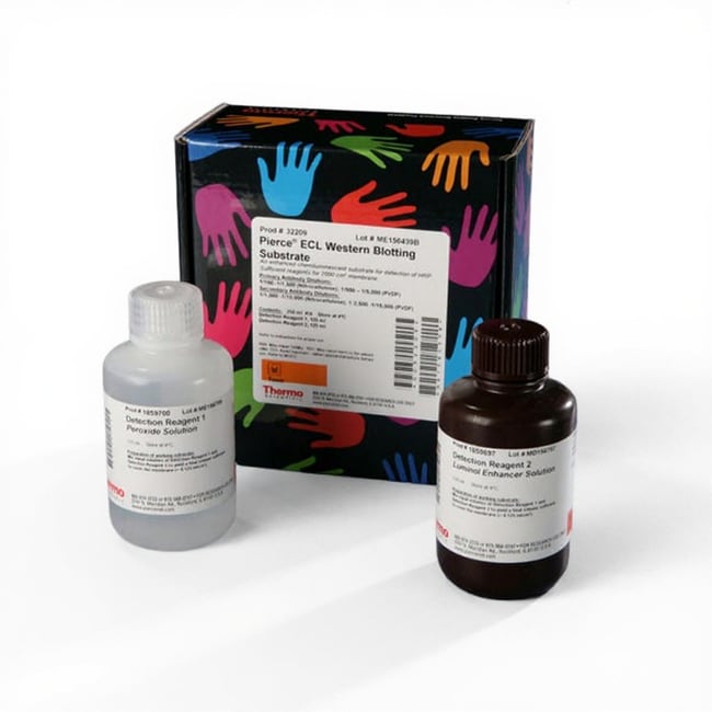


Thermo Scientific™ Pierce™ ECL Western Blotting Substrate
Description
Thermo Scientific Pierce ECL Western Blotting Substrate is a value-priced, entry-level horseradish peroxidase (HRP) substrate for enhanced chemiluminescence (ECL) that directly replaces costlier products without the need to re-optimize conditions.
Pierce ECL Plus Western Blotting Substrate characteristics include:
- Sensitivity: low picogram
- Stability: 1-hr working solution stability, 1-year kit stability at 4°C
- Compatibility: nitrocellulose and PVDF membranes
- Signal duration: 1 to 2 hours
- Recommended primary antibody concentration: 1:1,000–1:5,000 dilution (0.2–10 μg/mL)
- Recommended secondary antibody concentration: 1:1,000–1:15,000 dilution (0.07–1.0 μg/mL)
Pierce ECL Western Blotting Substrate provides reliability and performance equivalent to other standard ECL substrates for detection of HRP enzyme activity. It enables the detection of picogram amounts of antigen and allows for easy detection of HRP using photographic or other imaging methods.
Because the luminol and peroxide reagent formulations are identical to other commercially available substrate products, one can switch to Pierce ECL without needing to optimize probing conditions or incubation protocols. Pierce ECL Western Blotting Substrate provides performance identical to the original Amersham ECL Western Blotting Detection Reagent from GE Healthcare.
Specifications
Specifications
| Product Type | Substrate |
| Detection Method | Chemiluminescence |
| Form | Liquid |
| For Use With (Application) | Western Blot |
| Membrane Compatibility | Nitrocellulose, PVDF |
| Product Line | Pierce |
| Quantity | 250 mL |
| Substrate | Horseradish Peroxidase |
| Recommended Antibody Concentrations | 2°: 1:1-15K (0.07-1μg/mL), 1°: 1:1K (0.2- 10μg/mL) |
| Sensitivity | Low Picogram |
| Show More |
Frequently Asked Questions (FAQs)
Consider transferring to a different membrane or using a different detection method. We have observed increased sensitivity when using PVDF membranes in place of nitrocellulose. On PVDF membranes, using as little as 1 µg of total rat brain protein, PKC can be detected with alkaline phosphatase-mediated chromogenic detection in some cases using affinity-purified antibodies at a concentration of 0.5 µg/mL. Detection sensitivity can also be increased by using chemiluminescent detection, especially when using a SuperSignal West enhanced chemiluminescence subtrate (https://www.thermofisher.com/us/en/home/life-science/protein-biology/protein-assays-analysis/western-blotting/detect-proteins-western-blot/western-blot-detection-reagents/detection-technologies-western-blotting/chemiluminescent-western-blot-detection/supersignal-chemiluminescent-substrates.html) such as SuperSignal West Pico PLUS, SuperSignal West Dura, or SuperSignal West Femto. The secondary antibody should be used as recommend by the manufacturer and optimized as needed.
For Research Use Only. Not for use in diagnostic procedures.