missing translation for 'onlineSavingsMsg'
Learn More
Learn More
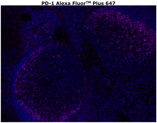
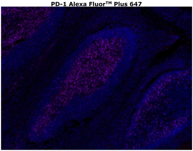
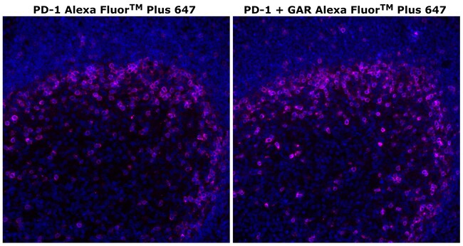
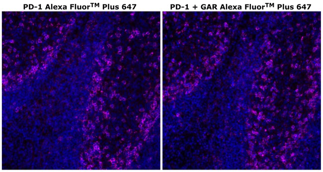
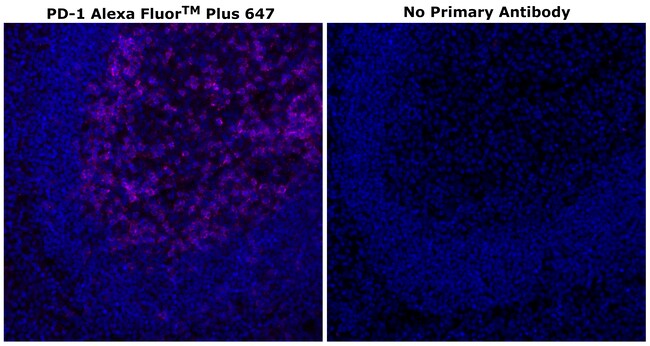
Invitrogen™ PD-1 (CD279) Recombinant Rabbit Monoclonal Antibody (RM309), Alexa Fluor™ Plus 647
Rabbit Recombinant Monoclonal Antibody
Supplier: Invitrogen™ 756800394
Description
Description: The RM309 antibody reacts with the human PD-1 (programmed death-1), a 55 kDa member of the immunoglobulin superfamily. PD-1 contains the immunoreceptor tyrosine-based inhibitory motif (ITIM) and plays a key role in peripheral tolerance and autoimmune disease. PD-1 is expressed predominantly on activated T and B lymphocytes. Two novel members of the B7 family have been identified as the PD-1 ligands, PD-L1 (B7-H1) and PD-L2 (B7-DC). Evidence reported to date suggests overlapping functions for these two PD-1 ligands and their constitutive expression on some normal tissues and upregulation on activated antigen-presenting cells. Applications Reported: This RM309 antibody has been reported for use in immunoblotting, immunohistochemical staining of formalin-fixed paraffin embedded tissue. Applications Tested: This RM309 antibody has been tested by immunohistochemistry on formalin-fixed paraffin embedded human tissue using high pH antigen retrieval and can be used at 3.3 μg/mL. It is recommended that the antibody be carefully titrated for optimal performance in the assay of interest. Using conjugate solutions: Centrifuge the protein conjugate solution briefly in a microcentrifuge before use; add only the supernatant to the experiment. This step will help eliminate any protein aggregates that may have formed during storage, thereby reducing nonspecific background staining.
Cell-mediated immune responses are initiated by T lymphocytes that are themselves stimulated by cognate peptides bound to MHC molecules on antig en-presenting cells (APC). T-cell activation is generally self-limited as activated T cells express receptors such as PD-1 (also known as PDCD-1) that mediate inhibitory signals from the APC. PD-1 can bind two different but related ligands, PDL-1 and PDL-2. Upon binding to either of these ligands, signals generated by PD-1 inhibit the activation of the immune response in the absence of 'danger signals' such as LPS or other molecules associated with bacteria or other pathogens. Evidence for this is seen in PD1-null mice who exhibit hyperactivated immune systems and autoimmune diseases. Despite its predicted molecular weight, PD-1 often migrates at higher molecular weight in SDS-PAGE.Specifications
| PD-1 (CD279) | |
| Recombinant Monoclonal | |
| 0.2 mg/mL | |
| PBS with 0.5% BSA, 10% proprietary stabilizer and 0.05% sodium azide; pH 7.2 | |
| Q15116 | |
| Pdcd1 | |
| A peptide corresponding to the N-terminus of human PD-1. | |
| 500 μL | |
| Primary | |
| Human | |
| Antibody | |
| IgG |
| Immunohistochemistry (Paraffin), Multiplex Immunohistochemistry | |
| RM309 | |
| Alexa Fluor Plus 647 | |
| Pdcd1 | |
| CD279; EGK_05005; hPD1; hPD-1; hPD-l; hSLE1; Ly101; mPD-1; PD1; PD-1; Pdc1; Pdcd1; programmed cell death 1; programmed cell death 1 protein; programmed cell death protein 1; programmed cell death protein 1-like; programmed death 1; Protein PD1; protein PD-1; sCD279; SLEB2; soluble CD279; systemic lupus erythematosus susceptibility 2 | |
| Rabbit | |
| Protein A | |
| RUO | |
| 5133 | |
| 4°C, store in dark | |
| Liquid |