Learn More

Invitrogen™ Low Fluorescence PVDF Membranes
Description
Invitrogen Low Fluorescence PVDF membranes are specifically optimized to provide exceptional performance in western blot, fluorescent total protein normalization, and dot blot applications. These membranes maintain high protein binding characteristics while providing minimal auto-fluorescence across a wide range of excitation wavelengths (280–800 nm). As a result, Low Fluorescence PVDF membranes provide excellent multi-plexing capability and help achieve higher signal-to-noise ratios and improved detection limits across a broad spectrum of fluorescent dyes.
Features and benefits
- Low background autofluorescence—reduced background noise across multiple wavelengths, providing superior signal-to-noise
- Consistency and reproducibility—optimized pore size engineered to produce exceptional results, regardless of protein size and abundance
- Versatile formats—assortment of sizes and formats to match specific laboratory needs
Find the perfect format
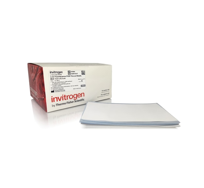
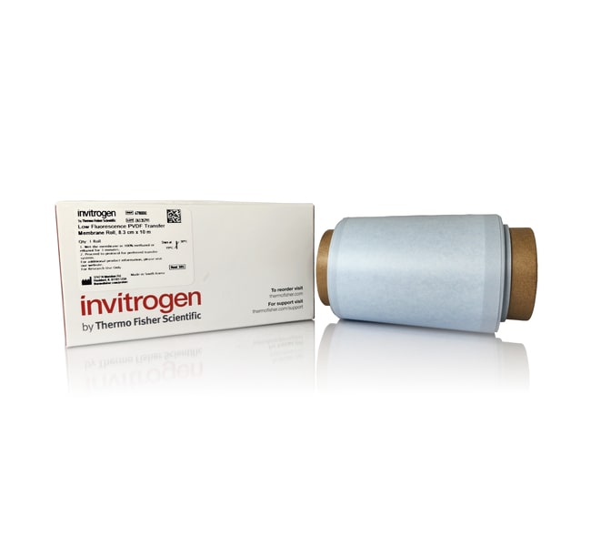
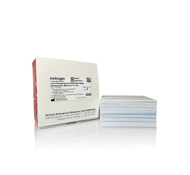
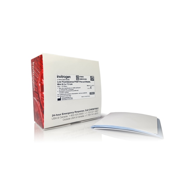
Low Fluorescence PVDF membranes are available in rolls, pre-cut sheets, and pre-assembled membrane and filter paper sandwiches. The single-gel width roll can be easily cut to length for use with either mini or midi gel transfers. Precut sizes are available for both mini and midi gels, reducing handling effort and saving time. Compatible with wet tank transfer and semi-dry transfer systems, Low Fluorescence PVDF membranes pair with any preferred protein gel, helping improve data quality without changing the workflow.
Optimized pore size
Low Fluorescence PVDF membranes are manufactured with a narrower distribution of pore sizes compared to conventional PVDF membranes. The pore distribution of our 0.3 μm Low Fluorescence PVDF membranes is optimized to deliver consistent and reproducible results for western blotting applications, irrespective of target protein size. Low Fluorescence PVDF membranes facilitate uniform protein transfer and detection, helping ensure high-quality, reliable western blot data.
Applications
- Fluorescent western blotting with IR and RGB fluorophores
- Total protein normalization and quantitative western blotting
- Chemiluminescent western blotting
Specifications
Specifications
| Length (Metric) | 13.5 cm |
| Width (Metric) | 8.3 cm |
| For Use With (Application) | Western Blotting |
| Quantity | 10/Box |
| Format | Sheet |
| Material | PVDF |
| Pore Size | 0.3 μm |
| Content And Storage | Store at room temperature. |
| Shipping Condition | Room temperature |
| Dimensions (LxW) | 13.5 x 8.3 cm |
| Show More |
Frequently Asked Questions (FAQs)
We recommend wetting PVDF in 100% methanol or ethanol for 3 min and then rinsing with deionized (DI) water before use. Wetting for shorter times can result in incomplete activation, leading to inconsistent protein binding. Wetting for a longer time will not have a negative impact.
PVDF is a hydrophobic membrane that will not readily interact or wet in water. To allow protein transfer and binding, the membrane must be initially wetted in methanol or ethanol and then rinsed with deionized (DI) water to allow protein binding to occur during transfer.
If the PVDF membrane dries out after transfer and before immunodetection, the membrane can be re-wetted by soaking the membrane in 100% methanol or ethanol for 3 min and then rinsing with deionized (DI) water before proceeding to the blocking step. This re-wetting will generally not negatively impact protein binding.
If the PVDF membrane dries out before imaging, for fluorescent western blots, it can typically be imaged dry without re-wetting. However, note that some fluorescent dyes are prone to degradation which can accelerate when the membrane is dry. If wetting is desired, soak the membrane in 100% methanol or ethanol for 3 min and then rinse with DI water and proceed to imaging.
The recommended wash condition after each step is: TBS or PBS with 0.05% Tween 20 for 5 min, repeated 3 times.
Yes. For best stripping and re-probing results, we recommend air drying the membrane after the transfer prior to immunoblotting (it will require rehydration in 100% methanol or ethanol prior to immunoblotting). Drying helps to fix the transferred proteins onto the membrane, helping ensure they remain immobilized and do not diffuse or wash away during subsequent steps. Drying the membrane can improve the binding efficiency of antibodies during the blocking and probing stages, resulting in better signal detection and stronger, clearer bands.
For Research Use Only. Not for use in diagnostic procedures.