missing translation for 'onlineSavingsMsg'
Learn More
Learn More
Invitrogen™ CD63 Monoclonal Antibody (H5C6), PE-eFluor™ 610, eBioscience™, Invitrogen™

Mouse Monoclonal Antibody
$598.00
Specifications
| Antigen | CD63 |
|---|---|
| Clone | H5C6 |
| Concentration | 5 μL/Test |
| Applications | Flow Cytometry |
| Classification | Monoclonal |

Description
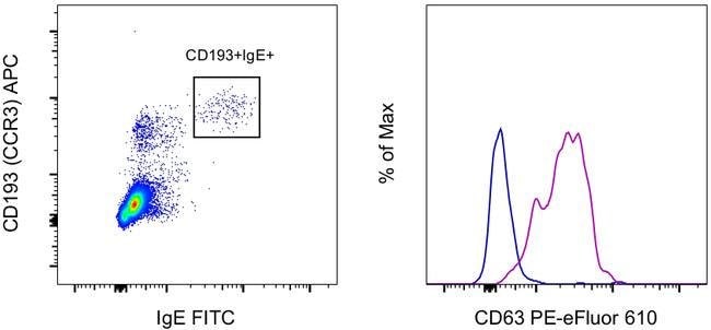
Description: This H5C6 monoclonal antibody reacts with human CD63, a type III member of the tetraspanin family of transmembrane proteins. CD63 is expressed intracellularly on lysosomes, endosomes, and granules of resting platelets and basophils. However, cell surface expression of CD63 can be detected on activated basophils and platelets, monocytes, macrophages, and granulocytes. This receptor is also expressed on endothelial cells, fibroblasts, and smooth muscle cells. Studies have demonstrated that CD63 associates with integrins (VLA-3 and VLA-6) and TIMP-1 to mediate the allergic response. Applications Reported: This H5C6 antibody has been reported for use in flow cytometric analysis. Applications Tested: This H5C6 antibody has been pre-diluted and tested by flow cytometric analysis of normal human peripheral blood cells. This may be used at 5 μL (1.0 μg) per test. A test is defined as the amount (μg) of antibody that will stain a cell sample in a final volume of 100 μL. Cell number should be determined empirically but can range from 10^5 to 10^8 cells/test. PE-eFluor 610 can be excited with laser lines from 488-561 nm and emits at 607 nm. We recommend using a 610/20 band pass filter (equivalent to PE-Texas Red). Please make sure that your instrument is capable of detecting this fluorochrome. Light sensitivity: This tandem dye is sensitive to photo-induced oxidation. Please protect this vial and stained samples from light.
CD63 (LAMP-3, lysosome-associated membrane protein-3), a glycoprotein of tetraspanin family, is present in late endosomes, lysosomes and secretory vesicles of various cell types. CD63 is also present in the plasma membrane, usually following cell activation. Hence, CD63 has become a widely used basophil activation marker. In mast cells, however, CD63 exposition does not need their activation. CD63 interacts with integrins and affects phagocytosis and cell migration, it is also involved in H/K-ATPase trafficking regulation of ROMK1 channels. CD63 also serves as a T-cell costimulation molecule. Expression of CD63 can be used for predicting the prognosis in earlier stages of carcinomas. CD63 is expressed on activated platelets, and is a lysosomal membrane glycoprotein that is translocated to plasma membrane after platelet activation. CD63 is also present in monocytes and macrophages and is weakly expressed on granulocytes, B, and T cells. CD63 is identical to the melanoma-associated antigen which is ME491 and to the platelet antigen PTLGP40. Diseases associated with CD63 dysfunction include melanoma and Hermansky-Pudlak Syndrome.Specifications
| CD63 | |
| 5 μL/Test | |
| Monoclonal | |
| Liquid | |
| RUO | |
| P08962 | |
| CD63 | |
| Primary | |
| 4°C, store in dark, DO NOT FREEZE! | |
| CD63 |
| H5C6 | |
| Flow Cytometry | |
| PE-eFluor 610 | |
| Mouse | |
| Human | |
| 967 | |
| IgG1 κ | |
| Affinity chromatography | |
| Antibody |