Learn More
Invitrogen™ EVOS™ Solution du package du système d’imagerie spatiale S1000
Description
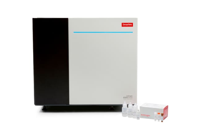
La Invitrogen EVOS solution du système d’imagerie spatiale S1000 comprend le système d’imagerie EVOS spatiale S1000, ainsi que des avantages de service supplémentaires et des réactifs d’imagerie spatiale pour assurer une expérience utilisateur plus complète.
Les ajouts incluent l’installation et la formation, une couverture de service prolongée, ainsi Aluora que le kit arc-en-ciel spatial avec ProLong verre (Cat. Non. A40002450). Ce kit contient tous les composants de réactifs nécessaires pour effectuer la détection d’amplification à base de tyramide 9-plex pour 100 diapositives.
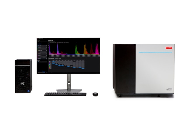
EVOS Système d’imagerie spatiale S1000
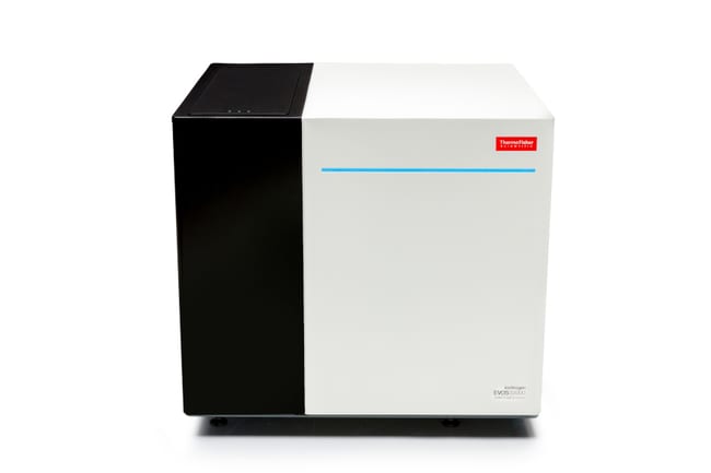
Le EVOS système d’imagerie spatiale S1000 est un instrument multimodal à haute performance pour l’imagerie et l’exploration de lames tissulaires utilisant les modes de fluorescence spectrale multiplex, champ clair transmis, contraste de phase et champ clair couleur. Les capacités spectrales du EVOS système S1000 permettent la capture et la résolution simultanées de jusqu’à huit cibles de fluorescence plus la coloration nucléaire (9-plex). Cela se fait en un seul cycle d’acquisition, évitant ainsi la nécessité de cycles répétés d’étiquetage et d’imagerie qui pourraient affecter l’intégrité tissulaire.
Le système EVOS S1000 propose une configuration matérielle hautement flexible comprenant un moteur d’éclairage à DEL personnalisé, une gamme étendue de filtres dichroïques et d’émission, ainsi qu’une caméra sCMOS haute sensibilité de 4,2 mégapixels (taille de pixel de 6,5 μm) permettant de détecter avec précision les architectures tissulaires les plus fines et complexes. Le système peut accueillir jusqu’à quatre lames simultanément et est livré avec trois objectifs préréglés (2,5X, 10X et 20X), avec la possibilité d’ajouter des objectifs apochromatiques supplémentaires de 5X et 40X afin d’étendre les capacités d’imagerie.
L’instrument est piloté par le logiciel EVOS S1000 Spatial, qui offre une interface avancée mais intuitive pour l’acquisition d’images de tissus entiers avec désenchevêtrement spectral intégré. Le logiciel permet des balayages d’aperçu rapides en modes champ clair transmis et fluorescence, ainsi qu’un mode « périscope » facilitant la navigation en temps réel dans les tissus. Il intègre également des fonctionnalités avancées d’autofocus permettant l’assemblage automatique des images et le désenchevêtrement spectral de lames complètes, éliminant ainsi la nécessité d’étapes de traitement post-acquisition hors ligne.
Le système EVOS S1000 Spatial Imaging offre les avantages clés suivants :
- Générateur de protocoles convivial permettant la sélection rapide des fluorophores via un menu déroulant, configurant automatiquement les paramètres optiques nécessaires pour atteindre le niveau de multiplexage souhaité
- Fonctions d’auto-exposition intuitive et d’autofocus puissant basé sur laser permettant de localiser rapidement et précisément les cibles d’intérêt
- Flux de travail guidé simple, cohérent et reproductible pour la résolution spectrale (désenchevêtrement linéaire) d’échantillons jusqu’à 9-plex à l’aide de contrôles non colorés et mono-couleur
- Protocoles prêts à l’emploi pour imagerie 9-plex et 7-plex, préconfigurés à partir de tissus de référence, permettant de démarrer rapidement l’analyse d’échantillons archivés
- Acquisition rapide et efficace d’images 9-plex sur zones personnalisées ou lames complètes, avec désenchevêtrement et assemblage réalisés automatiquement durant l’acquisition
- Production d’images OME-TIFF de haute qualité (résolution de 325 nm/pixel à 20X), déjà assemblées, désenchevêtrées et prêtes pour une analyse immédiate avec des logiciels tiers
- Compatibilité étendue avec des colorants fluorescents provenant de multiples fournisseurs (Alexa Fluor, Alexa Fluor Plus, Aluora Spatial Amplification, Opal), permettant l’utilisation de conjugués fluorescents ou de méthodes d’amplification spatiale
- Compatibilité complète avec des lames et lamelles standards, sans nécessité de consommables propriétaires
Imagerie spectrale simplifiée pour la biologie spatiale
Le système EVOS S1000 Spatial Imaging exploite la puissance de l’imagerie spectrale pour résoudre simultanément jusqu’à 9 fluorophores (8 cibles protéiques plus DAPI) en une seule acquisition. Il permet la sélection parmi plus de 30 fluorophores couvrant une plage d’émission de 450 à 810 nm. Les algorithmes intégrés de séparation spectrale, basés sur le désenchevêtrement linéaire, permettent une résolution claire et fiable des cibles protéiques sans recourir à des cycles répétés de marquage et d’imagerie, contribuant ainsi à préserver les échantillons biologiques précieux.
Matériel fiable, performant et évolutif
Le système comprend un porte-lames de précision à 4 positions monté sur une platine motorisée entièrement automatisée et contrôlée par logiciel. Il est capable de détecter plus de 60 combinaisons distinctes de longueurs d’onde d’excitation et d’émission, garantissant une évolutivité sans nécessité d’ajout de filtres supplémentaires pour de futurs projets de protéomique spatiale. Grâce à une capacité de 5 positions d’objectifs, les grossissements peuvent être configurés de 2,5X à 40X à l’aide d’objectifs de type apochromatique.
Une caméra sCMOS haute sensibilité (résolution 2040 × 2040 pixels) est utilisée pour capturer des images OME-TIFF monochromes 16 bits de haute qualité. Les images générées peuvent être enregistrées sur des disques internes (2 × 8 To SSD), des dispositifs USB externes ou des solutions infonuagiques approuvées. Un ordinateur externe Dell XE4 équipé d’un processeur Intel Core™ i9-12900 de 12e génération, de 128 Go de mémoire DDR4 et d’une carte graphique NVIDIA Quadro RTX™ A4000 assure le fonctionnement optimal du système.
Logiciel puissant conçu pour une simplicité avancée
Le logiciel EVOS S1000 Spatial permet aux utilisateurs d’accéder à l’imagerie multiplexée de manière simple et efficace. L’interface d’acquisition est conçue pour être intuitive, même pour les utilisateurs novices. Elle commence par un balayage d’aperçu extrêmement rapide pouvant être réalisé en mode transmis ou fluorescence. Par exemple, quatre lames peuvent être numérisées en fluorescence, y compris les marquages, en environ 180 secondes.
Le mode « périscope » en direct permet une navigation rapide dans les tissus. La localisation des échantillons est précise grâce à la combinaison d’un autofocus logiciel et d’un système de mise au point laser infrarouge. Un flux de travail guidé facilite la création de matrices de désenchevêtrement avec génération de rapports de qualité incluant des paramètres qualitatifs et quantitatifs pour évaluer la performance du processus.
Le logiciel permet également des acquisitions à grossissement variable pour champs de vision (FOV) et régions d’intérêt (ROI), avec des options d’automatisation avancées. L’assemblage des lames entières et le désenchevêtrement sont réalisés automatiquement dans le flux d’acquisition.
Préparation à l’analyse en aval
Les images acquises peuvent être analysées avec tout logiciel tiers compatible avec les fichiers OME-TIFF pyramidaux, facilitant la gestion de données d’imagerie multidimensionnelles volumineuses.
Composants supplémentaires inclus
La solution EVOS S1000 comprend l’installation et la formation assurées par les équipes de service et d’application. En plus de la garantie standard d’un an, une année supplémentaire de couverture est incluse, ainsi qu’une maintenance planifiée.
Le système inclut également le kit Aluora Spatial Rainbow avec ProLong Glass (Cat. No. A40002450), fournissant tous les réactifs nécessaires pour la détection 9-plex par amplification à la tyramide, incluant les colorants Aluora ainsi que les réactifs secondaires 4X GAM et 4X GAR, pour l’analyse d’environ 100 lames.
Spécifications
Spécifications
| Catégorie | Système d’imagerie configuré |
| Gamme de produit | EVOS |
| Caméra | Caméra sCMOS hautement sensible de 4,2 MP |
| À utiliser avec (application) | Fluorescence, champ clair, contraste de phase et imagerie couleur des lames tissulaires |
| À utiliser avec (équipement) | EVOS |
| Compatibilité à haut débit | Non |
| Source lumineuse | Moteur DEL personnalisé avec les spectres d’excitation suivants : 375 nm, 405 nm, 440 nm, 500 nm, 530 nm, 570 nm, 630 nm et 730 nm |
| Objectifs | Automatisé à 5 positions (incluant 2,5x, 10x et 20x) |
| Taille en pixels | 6,5 μm |
| Résolution | 2040 x 2040, 4.2 MP |
| Afficher plus de résultats |
Usage exclusivement réservé à la recherche. Ne pas utiliser dans des procédures de diagnostic.